Transmission Electron Microscopy
Preparation Procedure


              1. Specimen Preparation
              2. Viewing Preparation


FIXATION

     Fixation of tissues begins by mincing into 2-3 mm pieces and immersing them in Karnovsky's solution for 30 minutes, dicing further into smaller blocks of 0.5-1 mm on a side and continued fixation for an additional 2-5 hours. The specimens then undergo three 30 minute washes in cold 0.1M cacodylate buffer, with at least one overnight submersion. Tissue is then post-fixed for 2 hours in 1.33% ossmium tetroxide buffered with 0.1M cacodylate buffer. The tissue is again immersed in buffer solution for three 15 minute washes with a final rinse lasting 12 hours.

DEHYDRATION

     Dehydration of the tissue follows a series of alcohol washes at room temperature.

AlcoholTime
50% alcohol15 minutes
75% alcohol15 minutes
95% alcohol15 minutes
100% alcohol 30 minutes
Propylene Oxide 30 minutes


INFILTRATION

     In this stage, the general procedure is to expose the tissue to one or more mixtures of propylene oxide and embedding medium. Part one: preparation of the appropriate number of labeled molds dried in the oven. Part two: preparation of the infiltration resin, which is a complete mix of LX-112 embedding medium. Part three: after the second 30 minute rinse in propylene oxide, the following mixtures are prepared and used to infiltrate the tissue.

MixtureTime
Propylene Oxide/resin 2:12 hours
Propylene Oxide/resin 1:2Overnight
Propylene Oxide/resin 1:22 hours


EMBEDDING

     After preparing embedding mixture, molds are retrieved from the oven. Tissue is placed in a small drop of resin mix into the bottom of each mold. The molds are then filled to the top with resin. After all tissue is embedded, the molds are cured in a 45 degree oven for 8-20 hours followed by 24 hours in a 60 degree oven.


TRIMMING

     After molds are cured, the specimens are rough trimmed. The resin block is placed in a vice and trimmed by shaving the resin blocks into a trapezoid having the dimensions not exceeding 0.5 mm across the base, 0.4 mm across the top, and 0.3 mm along the sides.

SECTIONING

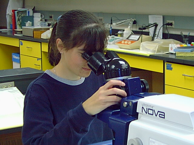
Ultratome in Use
     Sectioning is accomplished with the use of a Nova Ultratome. Sections of the trapezoids come off the knife and are floated onto water in the boat of a glass knife. Once trapezoids of gold or silver are available, a chloroformed Q-tip is wafted above them to expand the tissue; the tissue is then collected on 300 mesh copper grids.

STAINING

     Grids are stained in two steps. After covering a petri-dish containing a few NaOH pellets and a small sheet of dental wax, several drops of uranyl acetate stain are placed on the wax. The grids, with the specimen side down, remain in uranyl acetate for 10 minutes and are then rinsed in a series of four beakers of pure water. After rinsing, the grids are then stained with lead citrate for 15 minutes, rinsed again in pure water, and stored in a grid box.

After drying, the grids can be viewed.
To see the results of this long process, please check out the
TEM Student Micrograph Gallery.


Back to TEM Page